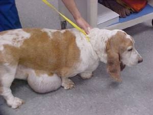
This dog has a large lipoma on the belly that was removed. Photo by Dr. Diane Sullivan
The nice thing about an aspirate is that it’s simple and inexpensive: just about every vet can do it. The downside is that some masses don’t shed cells easily, and all you have to look at are some cells on a slide. Sometimes that’s enough (as with lipomas) but sometimes you need a hunk of tissue to slice thinly to look at the arrangement of cells under a higher-powered microscope, which usually involves sending the sample to a pathologist. That chunk of tissue is a biopsy (no snowmen or toast involved), and it’s a surgical procedure that usually entails heavy sedation or anesthesia.
Performing an aspirate is the preferred way to diagnose a lipoma. They are usually quite easy to hit with a needle, and an aspirate is quick, painless and doesn’t require sedation or anesthesia. Most lipomas will shed cells easily and most vets who have ever worked a microscope can diagnose them right in the clinic with no need to get a pathologist involved. It’s usually a slam dunk.
Lipomas may look bad, but in most cases they’re unlikely to cause problems.
When can a lipoma cause a problem? We might recommend removal if it happens to be growing in a place that impedes getting around, or if it gets so big that carrying an extra few pounds of fat becomes hard to do. I’ve seen several large masses – the size of my head – that grew in the armpit or groin, and dogs are literally tripping over them. They can become so big they need to be removed just so a dog can walk. Those are a rarity, but they can happen.
I had a lipoma, too.
It wasn’t a big one, but it was big enough that I had a noticeable lump on my back [insert appropriate Quasimodo joke here]. I asked my GP about taking it off and he palpated it a bit and said “Sure, I can do it.”
He was halfway into it (he used local anesthesia and gave me some leather to bite on) when he said: “I think I’m in over my head here.” Not fun to hear when your skin is open and blood is running into your armpit.
Nevertheless, he pressed on and removed it. He found the limits of where his local anesthesia was by cutting something, I’d scream, and that was our little code that I needed more local anesthetic.
He botched closing up the incision, too. He only closed the skin layer, a mistake any first-year med student would’ve caught. I developed a big pocket of fluid called a seroma and had to have a drain placed in it. (Normally you close something like that in three layers; I couldn’t see what he was doing, so I didn’t catch it either.) I was a gooey, oozing mess for a couple of weeks, like a walking plate of nachos.
I still have a lovely scar; I tell people it is either from a bar fight or from that time I got shanked with a shiv in prison. (It depends on my mood.)
My non-medical mom called after the procedure and asked “How’s your lymphoma?” Lipoma, mom. Lipoma.
In any case, most lipomas in dogs are nothing more than a minor cosmetic problem. Make sure your vet sticks a needle in it to confirm its benign nature and aspirate or biopsy any other lumps that are suspicious. Cancer can look just like a lipoma, so better to be safe than sorry and don’t fall into the trap of thinking it looks benign – your eyes aren’t microscopes.
And please keep an eye on those little lipoma suckers; they can grow and cause problems if they’re in a high-motion area. It’s far easier to take them off when they’re small. And tell your vet to remember to close the skin in three layers while you’re at it. Or don’t and maybe your dog and I can compare scars.